🎬 Video Summary
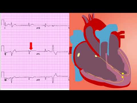
This video provides a concise explanation of how to read an ECG (Electrocardiogram) specifically in the context of Ischemic Heart Disease. Ischemic Heart Disease, a condition involving reduced blood flow to the heart muscle, can be diagnosed and monitored effectively with ECG interpretation. This guide offers practical insights for healthcare professionals and students to improve their diagnostic skills and patient care related to heart health.
🧠Teaching Pearls
- Learn to identify key ECG changes indicative of myocardial ischemia, such as ST-segment elevation or depression.
- Understand how different ECG leads correlate with specific regions of the heart affected by ischemia.
- Differentiate between acute and chronic ischemic changes on an ECG tracing.
- Recognize common ECG patterns associated with different types of Ischemic Heart Disease.
- Enhance your ability to quickly assess ECGs in emergency situations for timely intervention.
❓ Frequently Asked Questions
Q: What are the earliest ECG changes in myocardial ischemia?
A: The earliest ECG changes often involve T-wave inversion or hyperacute T-waves, followed by ST-segment elevation or depression depending on the type and location of ischemia.
Q: How accurate is ECG in diagnosing Ischemic Heart Disease?
A: While ECG is a valuable tool, its accuracy can vary. It’s highly specific but may have lower sensitivity, especially in early or mild cases. Other diagnostic tests may be required.
Q: Can an ECG detect all types of Ischemic Heart Disease?
A: ECG is most effective in detecting acute ischemic events like ST-segment elevation myocardial infarction (STEMI). It may be less sensitive for stable angina or non-ST-segment elevation myocardial infarction (NSTEMI).
Q: What are the limitations of using ECG for diagnosing Ischemic Heart Disease?
A: ECG limitations include its inability to detect small areas of ischemia, the presence of confounding factors (like electrolyte imbalances), and the potential for false negatives or false positives.
Q: How often should an ECG be performed for patients at risk of Ischemic Heart Disease?
A: The frequency of ECG monitoring depends on the individual’s risk factors and clinical presentation. In acute situations, serial ECGs may be necessary, while stable patients might require less frequent monitoring.
Q: What other tests complement ECG in diagnosing Ischemic Heart Disease?
A: Besides ECG, other tests like cardiac biomarkers (troponin), echocardiography, stress tests, and coronary angiography may be used to confirm and assess the severity of Ischemic Heart Disease.
🧠 Key Takeaways
- 💡 Understanding of ECG criteria for identifying myocardial ischemia in various clinical scenarios.
- 💡 Ability to correlate ECG findings with specific anatomical regions of the heart affected by ischemia.
- 💡 Knowledge of different ECG patterns associated with acute coronary syndromes and stable angina.
- 💡 Improved skill in interpreting ECG changes indicative of reversible and irreversible myocardial damage.
- 💡 Increased confidence in utilizing ECG as a crucial diagnostic tool in managing patients with Ischemic Heart Disease.
🔍 SEO Keywords
ECG interpretation, Ischemic Heart Disease, myocardial ischemia, ECG changes in ischemia, STEMI diagnosis, NSTEMI diagnosis, cardiac ECG reading.
“`