🎬 Video Summary
Learn to identify potassium abnormalities on an ECG with our comprehensive guide. This video breaks down how changes in potassium levels, from hypokalemia to hyperkalemia, manifest on an electrocardiogram, enabling you to quickly recognize and understand these critical cardiac indicators. Master ECG interpretation and improve your diagnostic skills for potassium-related heart conditions.
🧠Teaching Pearls
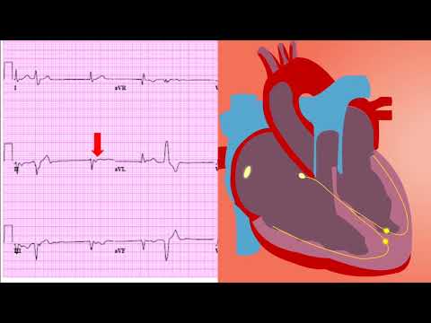
- 💡 Recognizing peaked T waves can be an early indicator of hyperkalemia on an ECG.
- 💡 U waves are often a telltale sign of hypokalemia, particularly when accompanied by flattened T waves.
- 💡 Prolongation of the PR interval can indicate hyperkalemia, affecting the heart’s conduction system.
- 💡 Understand the sequential ECG changes associated with worsening hyperkalemia for timely intervention.
- 💡 Always correlate ECG findings with the patient’s clinical presentation and potassium lab values.
❓ Frequently Asked Questions
Q: What ECG changes are associated with hyperkalemia?
A: Hyperkalemia can cause peaked T waves, widened QRS complexes, prolonged PR intervals, and ultimately, loss of P waves and a sine wave appearance on the ECG.
Q: How does hypokalemia affect the ECG?
A: Hypokalemia often leads to flattened or inverted T waves, prominent U waves, and ST segment depression on the ECG.
Q: Can an ECG accurately diagnose potassium imbalances?
A: An ECG can suggest potassium imbalances, but it should always be confirmed with a serum potassium level. The ECG changes can guide clinical suspicion.
Q: What is the significance of peaked T waves on an ECG?
A: Peaked T waves are an early sign of hyperkalemia, particularly in the setting of renal failure or medications that affect potassium levels.
Q: How quickly can ECG changes occur with severe hyperkalemia?
A: ECG changes can occur rapidly with severe hyperkalemia, sometimes within minutes, highlighting the need for prompt recognition and treatment.
Q: What other electrolyte imbalances can mimic potassium abnormality ECG changes?
A: While characteristic, electrolyte abnormalities like hypocalcemia and hypercalcemia can also cause ECG changes, emphasizing the need for a complete workup.
🧠 Key Takeaways
- 💡 Recognize the specific ECG patterns associated with both hypokalemia (U waves, flattened T waves) and hyperkalemia (peaked T waves, widened QRS).
- 💡 Understand the progressive ECG changes that occur as potassium levels fluctuate outside the normal range.
- 💡 Learn to differentiate between the ECG manifestations of mild, moderate, and severe potassium imbalances.
- 💡 Appreciate the clinical importance of correlating ECG findings with the patient’s history, symptoms, and lab results.
🔍 SEO Keywords
ECG interpretation, hyperkalemia ECG, hypokalemia ECG, potassium abnormalities, peaked T waves, U waves ECG, electrolyte imbalance ECG.
“`