🎬 Video Summary
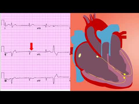
In Day 5 of the 30-day ECG Challenge, we demystify T waves! This video provides a comprehensive overview of T wave morphology, interpretation, and clinical significance. Whether you’re a medical student, nurse, or seasoned cardiologist, this guide will enhance your ECG reading skills and understanding of cardiac electrophysiology, specifically focusing on T wave analysis.
🧠 Teaching Pearls
- 💡 Understand the normal morphology and characteristics of T waves on an ECG.
- 💡 Learn to identify common T wave abnormalities, such as inversion, flattening, and hyperacute T waves.
- 💡 Explore the clinical significance of T wave changes in various cardiac conditions, including ischemia and electrolyte imbalances.
- 💡 Differentiate between benign and pathological T wave variations.
- 💡 Improve your ECG interpretation skills by focusing on the T wave in the context of the entire ECG tracing.
❓ Frequently Asked Questions
Q: What does an inverted T wave indicate?
A: Inverted T waves can indicate myocardial ischemia, old myocardial infarction, or non-specific repolarization abnormalities. The clinical context and other ECG findings are crucial for accurate interpretation.
Q: What are hyperacute T waves, and why are they important?
A: Hyperacute T waves are tall, broad T waves that can be an early sign of acute myocardial infarction. Recognizing them promptly is essential for timely intervention and improved patient outcomes.
Q: How do electrolyte imbalances affect T waves?
A: Electrolyte imbalances, such as hyperkalemia and hypokalemia, can significantly alter T wave morphology. Hyperkalemia often causes peaked T waves, while hypokalemia can lead to flattened or inverted T waves.
Q: Can medication affect T waves?
A: Yes, certain medications, like digoxin and some antiarrhythmics, can affect T wave morphology. Understanding these drug effects is crucial for accurate ECG interpretation.
Q: What’s the difference between a normal variant and a pathological T wave?
A: Normal variant T waves are benign findings that do not indicate underlying cardiac disease. Pathological T waves, on the other hand, are associated with cardiac abnormalities and require further investigation. Clinical history and serial ECGs can help differentiate between the two.
Q: How should I approach analyzing T waves on an ECG?
A: When analyzing T waves, consider their amplitude, morphology, direction, and relationship to other ECG components like the QRS complex and ST segment. Always interpret T waves in the context of the patient’s clinical presentation.
🧠 Key Takeaways
- 💡 Recognize normal T wave morphology and variations.
- 💡 Identify and interpret common T wave abnormalities.
- 💡 Understand the clinical significance of T wave changes in various cardiac conditions.
- 💡 Correlate T wave findings with patient history and other ECG components.
- 💡 Enhance your ECG interpretation skills for better patient care.
🔍 SEO Keywords
ECG interpretation, T wave abnormalities, cardiac electrophysiology, myocardial ischemia, hyperacute T waves, electrolyte imbalances, 30-day ECG challenge.
“`