case_study
Case Study: Early Repolarization in ECG of a Young Athlete
Identifying benign ECG patterns to reduce unnecessary workup
By Dr. Raj K | Published on June 21, 2025
Background
Early repolarization is a common ECG pattern seen in up to 10% of healthy young athletes. Although traditionally considered benign, it has occasionally been associated with idiopathic ventricular arrhythmias. Therefore, identifying it correctly can help avoid unnecessary investigations and anxiety.
Case Presentation
A 20-year-old male college-level basketball player presented for a preseason cardiovascular screening. He was asymptomatic with no history of chest pain, syncope, or family history of sudden death.
Physical Examination: Normal vital signs and cardiovascular exam
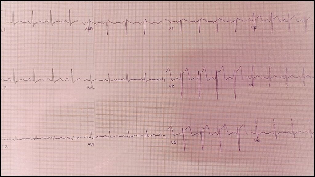
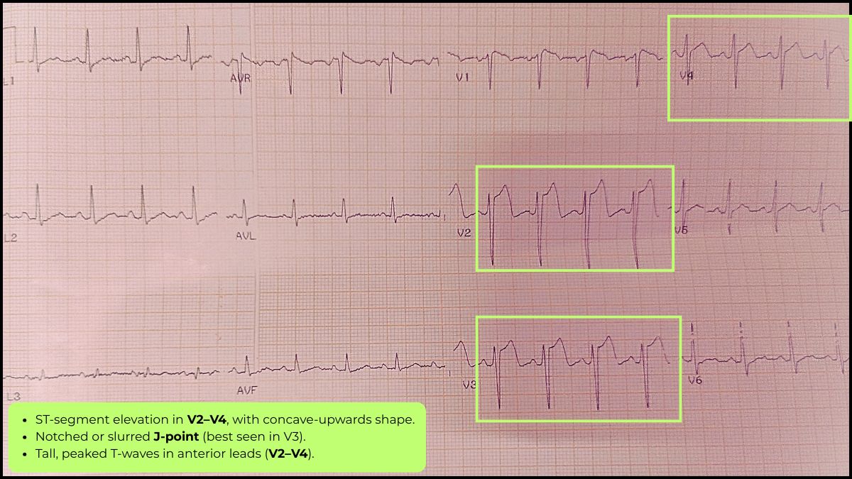
ECG Findings:
- J-point elevation of 1.5 mm in leads V2–V5
- Tall, peaked T waves
- No reciprocal changes, Q waves, or ST-depression
These features aligned well with benign early repolarization.
Discussion
Benign early repolarization typically features:
- J-point elevation ≥ 0.1 mV in 2 contiguous leads (mostly precordial leads V2–V5)
- Upsloping ST segments
- Symmetric T waves
Differentiate from ischemia using these red flags:
- Horizontal or down-sloping ST elevation
- Reciprocal ST depression
- Presence of Q waves
- Clinical symptoms (e.g., chest pain)
When benign features are confirmed in asymptomatic individuals, no further workup is required.
Key Takeaways
- Early repolarization is common and typically benign in young athletes
- Always assess ECG in clinical context — symptoms and exam matter
- Recognizing benign patterns reduces unnecessary referrals and tests
Gussak I, Antzelevitch C. Early repolarization syndrome: clinical characteristics and possible cellular and ionic mechanisms. J Electrocardiol. 2000 Oct;33(4):299-309. doi:10.1054/jelc.2000.18106. PMID: 11099355.
Related ECG News
-
Wearable ECG Tech Spots Post-Surgery Heart Risks: Vivalink & Brigham Study Paves The Way For Safer Recoveries
July 15, 2025 -
NIT‑Rourkela’s New Atrial Lead System Enhances ECG Clarity for Arrhythmia Detection
July 13, 2025 -
AI-Enhanced ECG May Help Detect Cognitive Decline Years Before Symptoms Appear, AHA Study Finds
July 13, 2025