🎬 Video Summary
Dive into Day 6 of our ECG challenge as we unravel the complexities of pathological Q waves. This video provides a concise and insightful exploration of identifying and interpreting these significant ECG abnormalities. Perfect for medical students, nurses, and practicing clinicians looking to enhance their ECG reading skills, this episode focuses on diagnosing myocardial infarctions and other cardiac conditions.
🧠 Teaching Pearls
- 💡 Pathological Q waves are significant indicators of prior myocardial infarction (MI).
- 💡 Differentiating between normal and pathological Q waves is crucial for accurate ECG interpretation.
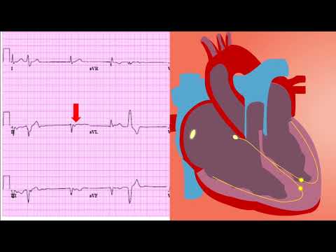
- 💡 Q wave morphology, including width and depth, helps determine its pathological significance.
- 💡 Location of pathological Q waves provides clues about the affected area of the heart.
- 💡 Consider clinical context alongside ECG findings for comprehensive patient assessment.
❓ Frequently Asked Questions
Q: How do I differentiate between normal and pathological Q waves?
A: Pathological Q waves are typically wider (≥0.04 seconds) and deeper (≥1/3 of the R wave amplitude) than normal Q waves. Their presence in leads where they are not normally seen is also a key indicator.
Q: What does a pathological Q wave indicate on an ECG?
A: The most common cause of pathological Q waves is prior myocardial infarction (MI), indicating dead or non-functional myocardial tissue. They can also be seen in other conditions such as hypertrophic cardiomyopathy or infiltrative myocardial diseases.
Q: Are pathological Q waves always indicative of a previous heart attack?
A: While myocardial infarction is the most common cause, pathological Q waves can also be present in other conditions, including left ventricular hypertrophy, Wolff-Parkinson-White syndrome, and certain cardiomyopathies. Clinical correlation is crucial.
Q: What is the significance of Q wave location in an ECG?
A: The location of pathological Q waves can help identify the specific area of the heart affected by infarction. For example, Q waves in leads II, III, and aVF suggest an inferior MI, while Q waves in V1-V4 suggest an anterior MI.
Q: How soon after a myocardial infarction do pathological Q waves appear on an ECG?
A: Pathological Q waves may not appear immediately after an MI. They typically develop over hours to days and can be a permanent feature on the ECG, representing the scar tissue from the infarction.
Q: Can pathological Q waves disappear over time?
A: In some cases, pathological Q waves may regress or diminish over time, particularly if there has been successful reperfusion therapy. However, in many cases, they remain as a permanent ECG marker of a prior MI.
🧠 Key Takeaways
- 💡 Learn to recognize the criteria for pathological Q waves on an ECG.
- 💡 Understand the clinical significance of pathological Q waves in diagnosing myocardial infarction.
- 💡 Identify the location of myocardial damage based on Q wave distribution.
- 💡 Differentiate pathological Q waves from normal Q waves and other ECG abnormalities.
- 💡 Integrate ECG findings with patient history and other diagnostic data for accurate interpretation.
🔍 SEO Keywords
Pathological Q waves, ECG interpretation, Myocardial infarction, ECG challenge, Cardiac diagnosis, ECG abnormalities, Heart attack ECG
“`