🎬 Video Summary
This video provides a concise overview of ECG features associated with pericardial effusion, with a special focus on electrical alternans. Learn to identify key ECG patterns and improve your interpretation skills for diagnosing this important cardiac condition. This tutorial is perfect for medical students, residents, and healthcare professionals seeking to enhance their knowledge of pericardial effusion and ECG analysis.
🧠 Teaching Pearls
- Understand the characteristic ECG changes seen in pericardial effusion.
- Learn to recognize electrical alternans and its significance.
- Differentiate between various ECG patterns associated with pericardial effusion.
- Improve your skills in interpreting ECGs for accurate diagnosis.
- Gain insights into the underlying mechanisms causing these ECG findings.
❓ Frequently Asked Questions
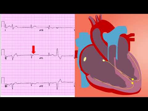
Q: What is electrical alternans, and why does it occur in pericardial effusion?
A: Electrical alternans refers to alternating amplitude changes in the QRS complex on the ECG. In pericardial effusion, this occurs due to the heart swinging within the fluid-filled pericardial sac, causing variations in the electrical axis.
Q: What are the most common ECG findings in pericardial effusion?
A: The most common findings include sinus tachycardia, low voltage QRS complexes, and, in some cases, electrical alternans.
Q: How can I differentiate pericardial effusion from other cardiac conditions using an ECG?
A: Look for the combination of low voltage, electrical alternans (if present), and clinical context. Echocardiography is often needed to confirm the diagnosis.
Q: Is electrical alternans always present in pericardial effusion?
A: No, electrical alternans is not always present. It is a specific but not a universally observed finding. Its presence strongly suggests the diagnosis.
Q: What is the significance of low voltage QRS complexes in pericardial effusion?
A: The fluid surrounding the heart attenuates the electrical signals, resulting in lower amplitude QRS complexes on the ECG.
Q: Can pericardial effusion be diagnosed solely based on ECG findings?
A: While ECG findings can be suggestive, echocardiography is the gold standard for confirming the diagnosis and assessing the size of the effusion.
🧠 Key Takeaways
- 💡 Recognize the significance of low voltage QRS complexes in the context of pericardial effusion.
- 💡 Identify electrical alternans as a specific ECG finding associated with significant pericardial effusion.
- 💡 Understand how the swinging motion of the heart within the pericardial sac causes electrical alternans.
- 💡 Correlate ECG findings with clinical presentation and other diagnostic modalities for accurate diagnosis.
- 💡 Apply this knowledge to improve the interpretation of ECGs in patients with suspected pericardial effusion.
🔍 SEO Keywords
Pericardial Effusion, ECG, Electrical Alternans, Low Voltage QRS, ECG Interpretation, Cardiac Diagnosis, Cardiology ECG Patterns.
“`