🎬 Video Summary
This video provides a concise overview of the ECG features associated with pulmonary embolism, a critical condition characterized by acute pulmonary hypertension. Understanding these ECG changes is crucial for early diagnosis and prompt treatment. The video highlights key indicators and explains how hypoxia and pulmonary hypoxic vasoconstriction can impact ECG readings in patients with pulmonary embolism.
🧠Teaching Pearls
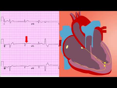
- 💡 Learn to recognize the classic S1Q3T3 pattern on an ECG, a possible indicator of pulmonary embolism.
- 💡 Understand how acute pulmonary hypertension affects the right ventricle and its reflection on the ECG.
- 💡 Be aware that hypoxia-induced pulmonary hypoxic vasoconstriction can also contribute to ECG changes in pulmonary embolism.
- 💡 Differentiate between various ECG abnormalities that may mimic pulmonary embolism to avoid misdiagnosis.
- 💡 Recognize that a normal ECG does not rule out pulmonary embolism, emphasizing the need for further investigation when clinically suspected.
❓ Frequently Asked Questions
Q: What is the most common ECG finding in pulmonary embolism?
A: While the S1Q3T3 pattern is often associated with pulmonary embolism, it’s not always present. Sinus tachycardia is the most common ECG finding, but it is non-specific.
Q: Can pulmonary embolism be ruled out based on a normal ECG?
A: No, a normal ECG cannot rule out pulmonary embolism. Further investigations, such as a CT pulmonary angiogram, are often needed to confirm or exclude the diagnosis.
Q: What does S1Q3T3 mean on an ECG?
A: S1Q3T3 refers to a deep S wave in lead I, a Q wave in lead III, and an inverted T wave in lead III on an ECG. It is suggestive of right ventricular strain.
Q: How does pulmonary hypertension affect the ECG in pulmonary embolism?
A: Pulmonary hypertension causes right ventricular strain, which can lead to right axis deviation, right bundle branch block, and T wave inversions in the anterior precordial leads on the ECG.
Q: Besides S1Q3T3, what other ECG changes might suggest pulmonary embolism?
A: Other ECG changes that might suggest pulmonary embolism include sinus tachycardia, right axis deviation, incomplete or complete right bundle branch block, T wave inversions in leads V1-V4, and atrial fibrillation.
Q: What is the significance of T wave inversions in pulmonary embolism?
A: T wave inversions, particularly in the anterior precordial leads (V1-V4), can indicate right ventricular strain or ischemia secondary to pulmonary embolism.
🧠 Key Takeaways
- 💡 Identify the S1Q3T3 pattern and its significance in the context of potential pulmonary embolism.
- 💡 Understand the relationship between pulmonary hypertension, hypoxia, and ECG changes in pulmonary embolism.
- 💡 Appreciate that ECG findings are often non-specific and should be interpreted in conjunction with clinical findings and other diagnostic tests.
- 💡 Recognize common ECG changes, such as sinus tachycardia and T wave inversions, that may be indicative of pulmonary embolism.
- 💡 Understand the limitations of ECG in diagnosing pulmonary embolism and when further investigation is warranted.
🔍 SEO Keywords
Pulmonary Embolism ECG, ECG findings in PE, S1Q3T3 pattern, Pulmonary Hypertension ECG, Hypoxia and ECG changes, ECG diagnosis of PE, Right ventricular strain ECG.
“`