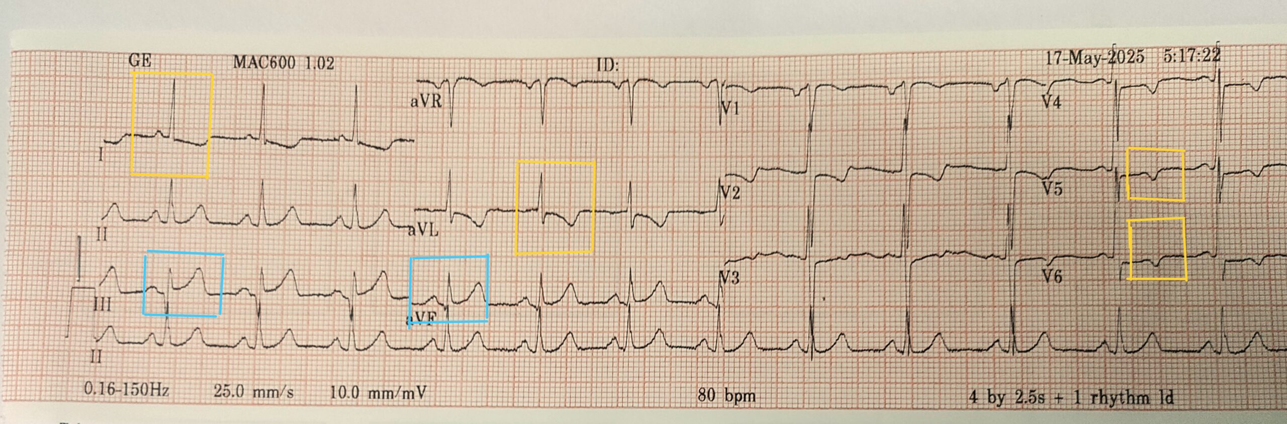
Acute Inferior STEMI with Lateral Ischemia
🏥 Clinical Context
A 54-year-old male presents with 45 minutes of crushing chest pain radiating to the jaw. History of type 2 diabetes and smoking. He is pale, tachypneic, and diaphoretic on arrival. Vitals: BP 96/60, HR 80 bpm, SpO₂ 92% RA.
🔍 Interpretation Steps
✅ Observations:
-
ST Elevation in Inferior Leads (II, III, aVF)
-
Clear upward deflection at J-point
-
Most prominent in III > II, which is classic for inferior STEMI
-
-
Reciprocal ST Depression in Lateral Leads (I, aVL, V5–V6)
-
ST segments are subtly but consistently depressed
-
Confirms posterior reciprocal changes
-
-
T Wave Inversions in V5–V6
-
Seen alongside ST depression
-
Sign of ischemia in lateral wall
-
-
Sinus rhythm ~80 bpm
-
Normal P waves and QRS morphology
-
QRS duration appears normal
-
📊 Final Diagnosis
Acute Inferior STEMI with Lateral Ischemia (reciprocal ST depression)
🎓 Teaching Pearl
Reciprocal ST depression in lateral leads confirms the diagnosis of an acute inferior MI and helps localize the ischemia zone more accurately.