🎬 Video Summary
This video provides a concise overview of the ECG features associated with an inferior wall myocardial infarction (MI). Learn how to identify key indicators on an ECG to quickly and accurately diagnose inferior wall MI. This educational resource is perfect for medical students, doctors, and healthcare professionals seeking to improve their ECG interpretation skills and patient care.
🧠Teaching Pearls
- 💡 Inferior wall MIs are often caused by occlusion of the right coronary artery (RCA) or the left circumflex artery (LCX).
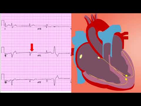
- 💡 Look for ST-segment elevation in leads II, III, and aVF to diagnose an inferior wall MI.
- 💡 Reciprocal ST-segment depression can be seen in the anterior leads (I, aVL, V1-V6).
- 💡 Be aware of right ventricular involvement in inferior wall MIs, as this impacts management.
- 💡 Early recognition and timely intervention are crucial for improving patient outcomes in inferior wall MIs.
❓ Frequently Asked Questions
Q: What ECG leads show ST elevation in inferior MI?
A: In an inferior wall MI, ST-segment elevation is typically seen in leads II, III, and aVF on the ECG.
Q: What is the most common cause of inferior wall MI?
A: The most common cause is occlusion of the right coronary artery (RCA), which supplies blood to the inferior wall of the heart.
Q: How can I differentiate inferior MI from pericarditis on an ECG?
A: While both can present with ST elevation, inferior MI usually shows reciprocal ST depression in anterior leads and may have Q waves, which are less common in pericarditis. Clinical context is also important.
Q: What are the initial steps in managing a patient with inferior wall MI?
A: Initial management includes oxygen administration, aspirin, nitroglycerin (if not contraindicated), morphine for pain, and prompt initiation of reperfusion therapy (PCI or thrombolytics).
Q: What are the complications associated with inferior wall MI?
A: Potential complications include heart block, right ventricular infarction, hypotension, and arrhythmias.
Q: Is an inferior MI always a STEMI?
A: Inferior MIs are often STEMIs (ST-Elevation Myocardial Infarctions) due to complete artery blockage, but can sometimes be NSTEMIs (Non-ST-Elevation Myocardial Infarctions) with partial blockage.
🧠 Key Takeaways
- 💡 Identify ST-segment elevation in leads II, III, and aVF to diagnose inferior wall MI.
- 💡 Recognize reciprocal ST-segment depression in anterior leads.
- 💡 Understand the importance of prompt diagnosis and reperfusion therapy.
- 💡 Be aware of potential complications, such as right ventricular infarction and heart block.
- 💡 Recognize the role of ECG interpretation in acute myocardial infarction management.
🔍 SEO Keywords
Inferior wall MI, ECG interpretation, ST-segment elevation, Myocardial infarction, Cardiology, Acute coronary syndrome, Medical education.
“`