🎬 Video Summary
This video provides a clear explanation of the ECG criteria used to diagnose left ventricular hypertrophy (LVH). Learn how to identify key indicators of LVH on an electrocardiogram and improve your diagnostic skills. This tutorial is perfect for medical students, residents, and practicing clinicians looking to enhance their understanding of ECG interpretation in the context of LVH.
🧠Teaching Perls
- 💡 Understand the specific voltage criteria used in ECG diagnosis of Left Ventricular Hypertrophy.
- 💡 Learn how to differentiate between various ECG patterns associated with LVH.
- 💡 Discover practical tips for accurately interpreting ECG results in the context of potential LVH.
- 💡 Familiarize yourself with the impact of LVH on the QRS complex and other ECG intervals.
- 💡 Explore the clinical significance of LVH and its implications for patient management.
❓ Frequently Asked Questions
Q: What are the main ECG criteria for Left Ventricular Hypertrophy?
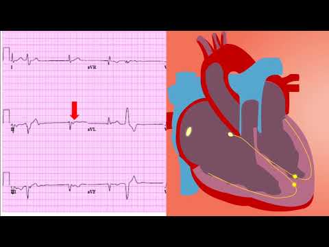
A: The main ECG criteria for LVH include increased QRS amplitude (S wave in V1 + R wave in V5 or V6 > 35 mm), ST-segment depression, and T-wave inversion in lateral leads.
Q: How accurate is ECG in diagnosing Left Ventricular Hypertrophy?
A: While ECG is a useful initial screening tool, its sensitivity in diagnosing LVH can vary. Echocardiography is often used for confirmation and more accurate assessment.
Q: What is the significance of ST-T wave changes in LVH?
A: ST-segment depression and T-wave inversion are common findings in LVH and can indicate repolarization abnormalities due to increased ventricular mass.
Q: Can hypertension cause Left Ventricular Hypertrophy?
A: Yes, chronic hypertension is a major cause of LVH as the heart has to work harder to pump blood against increased resistance.
Q: What is the role of echocardiography in diagnosing LVH?
A: Echocardiography is considered the gold standard for diagnosing LVH, as it allows direct measurement of left ventricular wall thickness and chamber size.
Q: Are there any specific ECG patterns in LVH with strain?
A: Yes, LVH with strain typically shows ST-segment depression and T-wave inversion in the lateral leads (V5, V6, I, aVL), indicating repolarization abnormalities.
🧠 Key Takeaways
- 💡 Identify the voltage criteria for diagnosing LVH on an ECG.
- 💡 Recognize ST-segment and T-wave changes associated with LVH.
- 💡 Understand the importance of considering clinical context when interpreting ECG findings for LVH.
- 💡 Learn how LVH can manifest differently on ECG depending on its underlying cause.
- 💡 Appreciate the limitations of ECG in diagnosing LVH and the role of other diagnostic modalities.
🔍 SEO Keywords
Left Ventricular Hypertrophy, LVH, ECG criteria, ECG diagnosis, Electrocardiogram, Hypertension, Cardiology, ECG interpretation
“`