🎬 Video Summary
In this insightful video, “Module 2: Mapping Heart Walls with ECG Leads | ECG Code Pro,” you’ll learn a critical skill for accurate ECG interpretation: identifying which ECG leads correspond to specific heart walls. Mastering this mapping technique is essential for diagnosing cardiac conditions and providing optimal patient care. This video provides clear instruction on ECG lead placement and correlation to heart anatomy.
🧠Teaching Pearls
- Gain a foundational understanding of ECG lead placement and their anatomical representation of the heart.
- Learn to correlate specific ECG lead changes with pathology in different heart walls.
- Discover techniques for confidently mapping ECG leads to anterior, inferior, lateral, and septal heart walls.
- Improve your diagnostic accuracy in identifying myocardial infarction and other ischemic events using ECGs.
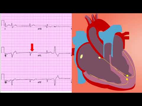
- Understand the significance of ECG lead groupings (e.g., inferior leads) for recognizing specific cardiac issues.
❓ Frequently Asked Questions
Q: How do I remember which ECG leads correspond to each heart wall?
A: A helpful mnemonic or visualization technique can aid in remembering the ECG lead-to-heart wall relationship. Focus on associating lead groupings (e.g., inferior leads II, III, aVF) with the corresponding anatomical location (inferior wall).
Q: What is the significance of reciprocal changes on an ECG?
A: Reciprocal changes, such as ST-depression in leads opposite the site of ST-elevation, can provide further evidence of myocardial ischemia or infarction and aid in localizing the affected heart wall.
Q: Which ECG leads are most important for assessing the anterior wall of the heart?
A: The anterior wall is primarily assessed using leads V1-V4. Changes in these leads should prompt a closer evaluation for anterior myocardial infarction.
Q: How accurate is ECG in detecting heart wall abnormalities?
A: ECG is a valuable tool but has limitations. While ST-elevation is a strong indicator of myocardial infarction, other factors and conditions can affect ECG findings. It is often used in conjunction with other diagnostic tools.
Q: What does lateral wall infarction look like on ECG?
A: Lateral wall infarctions are typically indicated by ST-elevation in leads I, aVL, V5, and V6.
Q: What is the best way to improve my ECG interpretation skills?
A: Consistent practice, review of ECG cases, and understanding the underlying cardiac anatomy and physiology are essential for improving your ECG interpretation skills. Seek out resources like ECG Code Pro for ongoing learning.
🧠 Key Takeaways
- 💡 You will be able to identify the anatomical area of the heart that each ECG lead represents.
- 💡 You will learn how to recognize patterns of ST-elevation or depression associated with specific heart wall infarctions.
- 💡 You will understand the importance of considering reciprocal changes in ECG interpretation.
- 💡 You will be able to use ECG lead mapping to improve diagnostic accuracy in suspected cardiac events.
🔍 SEO Keywords
ECG lead placement, heart wall mapping, ECG interpretation, anterior wall MI, inferior MI, lateral wall infarction, ECG diagnosis.
“`