🎬 Video Summary
In Module 3 of ECG Code Pro, dive into the essential foundations for ECG interpretation. This video comprehensively covers cardiac anatomy, coronary circulation, and the heart’s conduction system, providing crucial knowledge for understanding ECG readings. Learn the vital structures and pathways that influence heart function and electrical activity, laying the groundwork for confident ECG analysis.
🧠Teaching Pearls
- Gain a solid understanding of the heart’s anatomical structures and their relevance to ECG interpretation.
- Explore the intricacies of coronary circulation and its impact on myocardial health, crucial for identifying ischemic changes on an ECG.
- Master the pathway of the heart’s conduction system, from the SA node to the Purkinje fibers, to understand normal and abnormal rhythms.
- Learn how disruptions in any part of the cardiac anatomy, coronary circulation, or conduction system can manifest as specific ECG abnormalities.
- Develop a systematic approach to correlating anatomical knowledge with ECG findings for accurate diagnoses.
❓ Frequently Asked Questions
Q: What is the significance of the SA node in ECG interpretation?
A: The SA node is the heart’s natural pacemaker, initiating the electrical impulses that trigger each heartbeat. Its function directly influences the P wave on an ECG, so abnormalities in P wave morphology or rate can indicate SA node dysfunction.
Q: How does coronary artery disease affect ECG readings?
A: Coronary artery disease restricts blood flow to the heart muscle, potentially causing ischemia or infarction. These conditions can manifest on an ECG as ST-segment elevation or depression, T-wave inversion, or the presence of Q waves.
Q: What is the role of the AV node in regulating heart rhythm?
A: The AV node delays the electrical impulse from the atria to the ventricles, ensuring coordinated contraction. It also acts as a backup pacemaker if the SA node fails. Dysfunction in the AV node can lead to heart blocks, affecting the PR interval on an ECG.
Q: How does the Purkinje fiber network influence the QRS complex?
A: The Purkinje fibers rapidly conduct electrical impulses throughout the ventricles, triggering ventricular contraction. Their efficient function is crucial for a narrow QRS complex. Abnormalities in the Purkinje network can lead to widened QRS complexes, indicative of ventricular conduction delays.
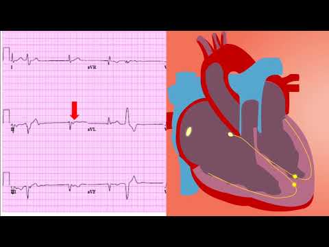
Q: What are the main coronary arteries, and how do they relate to ECG changes?
A: The main coronary arteries are the left anterior descending (LAD), the right coronary artery (RCA), and the left circumflex artery (LCX). Occlusion of each artery leads to specific ECG changes in related leads, such as ST-elevation MI (STEMI) patterns. For example, LAD occlusion often results in ST elevation in leads V1-V4.
Q: How does cardiac hypertrophy affect ECG readings?
A: Cardiac hypertrophy, or enlargement of the heart, can alter the electrical signals detected by an ECG. Left ventricular hypertrophy often presents with increased QRS amplitude and repolarization abnormalities, such as ST-segment depression and T-wave inversion, in the lateral leads.
🧠 Key Takeaways
- 💡 Understanding the heart’s anatomical structures provides a framework for interpreting ECG patterns.
- 💡 Coronary circulation knowledge is crucial for identifying ischemic changes and diagnosing coronary artery disease on an ECG.
- 💡 Familiarity with the conduction system helps in recognizing and classifying various arrhythmias.
- 💡 Correlating anatomical knowledge with ECG findings leads to more accurate diagnoses.
- 💡 Module 3 provides foundational knowledge for advanced ECG interpretation.
🔍 SEO Keywords
ECG interpretation, cardiac anatomy, coronary circulation, heart conduction system, ECG analysis, cardiac electrophysiology, ECG abnormalities
“`