🎬 Video Summary
This video provides a comprehensive overview of supraventricular tachycardia (SVT), a common type of heart rhythm disturbance. It delves into the different types of SVT and their characteristic ECG features, enabling healthcare professionals and students to accurately identify and differentiate between them. Learn to recognize key indicators and improve your diagnostic skills in managing patients with SVT.
🧠Teaching Pearls
- Understand the underlying mechanisms of different SVT types to improve diagnostic accuracy.
- Learn to differentiate between AV nodal reentrant tachycardia (AVNRT), AV reentrant tachycardia (AVRT), and atrial tachycardia on ECG.
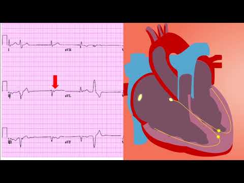
- Recognize specific ECG features such as P-wave morphology and RP intervals for accurate SVT classification.
- Improve your ability to identify subtle ECG patterns associated with different SVT subtypes.
- Master the interpretation of ECG findings crucial for guiding appropriate treatment strategies for SVT.
⏱ Timestamps
❓ Frequently Asked Questions
Q: What is the most common type of SVT?
A: AV nodal reentrant tachycardia (AVNRT) is the most common type of supraventricular tachycardia, characterized by a reentrant circuit within the AV node.
Q: How can I differentiate AVNRT from AVRT on an ECG?
A: In AVNRT, the P waves are often buried within the QRS complex or appear shortly after, resulting in a short RP interval. AVRT typically has a longer RP interval due to the accessory pathway.
Q: What are the key ECG features of atrial tachycardia?
A: Atrial tachycardia is characterized by distinct P waves preceding each QRS complex, with the P-wave morphology differing from the sinus P wave.
Q: What is the significance of the RP interval in SVT diagnosis?
A: The RP interval (from the onset of the QRS complex to the onset of the P wave) helps differentiate orthodromic (short RP) from antidromic (long RP) AVRT, as well as distinguish AVNRT from atrial tachycardia.
Q: What is the role of vagal maneuvers in SVT?
A: Vagal maneuvers, such as carotid sinus massage, can slow or terminate some SVTs, particularly AVNRT, by increasing vagal tone and affecting AV node conduction.
Q: What are the initial steps in managing a patient with SVT?
A: The initial steps include assessing the patient’s hemodynamic stability, obtaining an ECG, and considering vagal maneuvers or adenosine if appropriate. If unstable, synchronized cardioversion may be necessary.
🧠 Key Takeaways
- 💡 Learn the different types of supraventricular tachycardia (SVT) and their mechanisms.
- 💡 Identify characteristic ECG features that distinguish between AVNRT, AVRT, and atrial tachycardia.
- 💡 Understand the importance of P-wave morphology and RP intervals in SVT diagnosis.
- 💡 Recognize the role of vagal maneuvers and pharmacological interventions in SVT management.
- 💡 Improve your ability to interpret ECGs and manage patients presenting with SVT effectively.
🔍 SEO Keywords
Supraventricular tachycardia, SVT types, ECG features of SVT, AVNRT, AVRT, Atrial tachycardia, SVT diagnosis, ECG interpretation, cardiac arrhythmias.
“`